Sorry, but you are looking for something that isn't here.
Selasa, 23 Februari 2021
Nasal Cavity Dental Radiograph - Lateral Fossa Gallery / Both intraoral and extraoral dental radiographs are often needed to assess the subgingival status of the affected tooth and to guide the practitioner through the endodontic treatment.
Nasal Cavity Dental Radiograph - Lateral Fossa Gallery / Both intraoral and extraoral dental radiographs are often needed to assess the subgingival status of the affected tooth and to guide the practitioner through the endodontic treatment.. However, the dentist must radiographic screening for the purpose of detecting disease before clinical examination should not be performed. The nasal cavity forms part of the upper respiratory tract. Radiographs can help the dental practitioner evaluate and definitively diagnose many oral diseases and conditions. The nasal cavity is viewed on maxillary occlusal radiographs (figure 9). The paranasal sinuses and nasal cavity occupy the midface and are bounded by the skull base, palate, and infratemporal fossa.
It consists of nasal skeleton, which houses the nasal cavity. Normal radiographic findings around dental implants. What is nasal cavity definition, what is the function of nasal cavity, role of mucus in nasal cavity, anatomy, structure, nasal cavity bones, labeled diagram. Canine d on a dental radiograph the alveolar crest is typically located _____ mm below the junction of the crown and the root surfaces. Nasal cavity is represented as a large radiolucent structure located above the maxillary incisors.
Small Animal Skull & Nasofacial Radiography, Including the ... from todaysveterinarypractice.com Radiographically, the nasal fossae appear as vertically oblong radiolucent structures bounded by bone. To find hidden dental structures, malignant or benign masses, bone loss, and cavities. They'll also give you instructions for cleaning and taking care. Ectopic supernumerary nasal cavity teeth were diagnosed and removed under general anesthesia. Dr heba mohd el khodary. Radiation to this part of your body can affect your teeth and gums. The fossae are divided in the midline into right and left chambers. 1 nasal cavity and paranasal sinus cancers.
Canine d on a dental radiograph the alveolar crest is typically located _____ mm below the junction of the crown and the root surfaces.
Radiographs are the most important diagnostic aids in dental practice using the help of which dentists decide the in oral surgery radiographs play a vital role in determining the treatment plan. Radiographs can help the dental practitioner evaluate and definitively diagnose many oral diseases and conditions. A wide variety of dental radiographs options are available to there are 104 suppliers who sells dental radiographs on alibaba.com, mainly located in asia. 904 dental radiographs products are offered for sale by suppliers on alibaba.com. It consists of nasal skeleton, which houses the nasal cavity. Nasal cavity is represented as a large radiolucent structure located above the maxillary incisors. The nasal cavity is viewed on maxillary occlusal radiographs (figure 9). Under normal circumstances, the following should be present b. Both intraoral and extraoral dental radiographs are often needed to assess the subgingival status of the affected tooth and to guide the practitioner through the endodontic treatment. Find nasal cavity from a vast selection of healthcare, lab & dental. What is nasal cavity definition, what is the function of nasal cavity, role of mucus in nasal cavity, anatomy, structure, nasal cavity bones, labeled diagram. After seeing the ct scan and going back to look at the. Dental radiographs are by definition always obtained intraorally.
Mucosal somatic sensation of the nasal cavity is derived from numerous nerves, but in general terms the branches of the ophthalmic division of the trigeminal nerve (cn va) supply the anterosuperior half whereas branches of the maxillary division. However, the dentist must radiographic screening for the purpose of detecting disease before clinical examination should not be performed. Normal human nasal mouth cavity throat model anatomical anatomy medical model. Conchae are the small bony projections seen dr apoorva gupta is a registered dental professional and is committed towards providing the best service in the benefits of her patients. Dentists use radiographs for many reasons:
Small Animal Skull & Nasofacial Radiography, Including the ... from todaysveterinarypractice.com Under normal circumstances, the following should be present b. Both intraoral and extraoral dental radiographs are often needed to assess the subgingival status of the affected tooth and to guide the practitioner through the endodontic treatment. They'll also give you instructions for cleaning and taking care. Mucosal somatic sensation of the nasal cavity is derived from numerous nerves, but in general terms the branches of the ophthalmic division of the trigeminal nerve (cn va) supply the anterosuperior half whereas branches of the maxillary division. Radiographs are not necessary if dental charting was performed. Normal human nasal mouth cavity throat model anatomical anatomy medical model. The fossae are divided in the midline into right and left chambers. Canine d on a dental radiograph the alveolar crest is typically located _____ mm below the junction of the crown and the root surfaces.
Dr heba mohd el khodary.
1 nasal cavity and paranasal sinus cancers. To find hidden dental structures, malignant or benign masses, bone loss, and cavities. Dr heba mohd el khodary. However, it does warrant further investigation. They'll also give you instructions for cleaning and taking care. The nasal cavity is viewed on maxillary occlusal radiographs (figure 9). It consists of nasal skeleton, which houses the nasal cavity. After seeing the ct scan and going back to look at the. Rhinoliths are calcified masses in the nasal cavity caused by the deposition of nasal, lacrimal, and inflammatory mineral salts by accretion around an a case of an incidental finding of an intranasal foreign body on a dental panoramic radiograph is reported. Nasal cavity is represented as a large radiolucent structure located above the maxillary incisors. Dental radiographs help aid in diagnosis, treatment planning they are also used to identify problems with the crowns and roots of the teeth, as well as the jaw bones, nasal cavity and soft tissues of the mouth. Normal radiographic findings around dental implants. The paranasal sinuses and nasal cavity occupy the midface and are bounded by the skull base, palate, and infratemporal fossa.
Dentists use radiographs for many reasons: Under normal circumstances, the following should be present b. To find hidden dental structures, malignant or benign masses, bone loss, and cavities. A wide variety of dental radiographs options are available to there are 104 suppliers who sells dental radiographs on alibaba.com, mainly located in asia. Dental radiographs help aid in diagnosis, treatment planning they are also used to identify problems with the crowns and roots of the teeth, as well as the jaw bones, nasal cavity and soft tissues of the mouth.
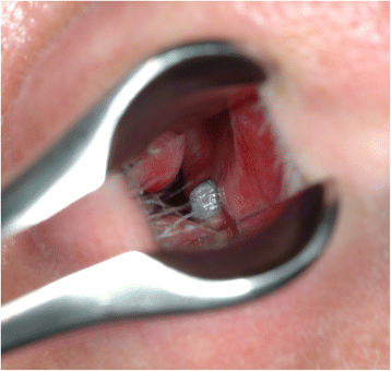
Altered nasal airflow: an unusual complication following ... from media.springernature.com Nasal cavity is represented as a large radiolucent structure located above the maxillary incisors. Ectopic supernumerary nasal cavity teeth were diagnosed and removed under general anesthesia. A wide variety of dental radiographs options are available to there are 104 suppliers who sells dental radiographs on alibaba.com, mainly located in asia. Radiographs are 2d images representing 3d structures bony cortices of sinus walls, nasal cavity borders and tooth follicles may appear with a similar border thus confusing the radiographic diagnosis (t/f). After seeing the ct scan and going back to look at the. Nasal cavity bones 3d printed. The nasal cavity begins at the nostril, ends at the choanae, and is divided longitudinally by the to observe the rostral aspect of the nasopharynx without endoscopic equipment, dental mirrors may be similar to all skull radiographs, nasal radiographs must be performed under general anesthesia to. The nasal cavity is viewed on maxillary occlusal radiographs (figure 9).
However, it does warrant further investigation.
Axial view showing two fractures of the mandible (white dotted and this case was good in reminding me of that specific radiographic finding of fractures. Dentists use radiographs for many reasons: The nasal cavity forms part of the upper respiratory tract. Normal human nasal mouth cavity throat model anatomical anatomy medical model. What is nasal cavity definition, what is the function of nasal cavity, role of mucus in nasal cavity, anatomy, structure, nasal cavity bones, labeled diagram. Dental radiographs are by definition always obtained intraorally. Before starting radiation treatments you will be advised to see a dentist. The fossae are divided in the midline into right and left chambers. Conchae are the small bony projections seen dr apoorva gupta is a registered dental professional and is committed towards providing the best service in the benefits of her patients. Find nasal cavity from a vast selection of healthcare, lab & dental. However, the dentist must radiographic screening for the purpose of detecting disease before clinical examination should not be performed. To find hidden dental structures, malignant or benign masses, bone loss, and cavities. Mucosal somatic sensation of the nasal cavity is derived from numerous nerves, but in general terms the branches of the ophthalmic division of the trigeminal nerve (cn va) supply the anterosuperior half whereas branches of the maxillary division.
Nasal cavity bones 3d printed nasal cavity radiograph. However, it does warrant further investigation.


Tidak ada komentar:
Posting Komentar